Make sure to sign up for an account today for exclusive coupons and free shipping on orders over $75!
Maximum quantity allowed is 999

Efficient organelle isolation is important for investigating protein localization and function. TCI offers kit products optimized for nuclear, cytoplasmic, and mitochondrial fractionation.
Mitochondrial Isolation Kit
Mitochondrial Isolation Kit (Product No. M3527) can be used to easily isolate mitochondria from cultured mammalian cells. Isolated mitochondria can be used for downstream analyses such as Western Blotting.
Kit Components
- Mitochondrial Isolation Reagent A for approx. 30 samples
- Mitochondrial Isolation Reagent B for approx. 30 samples
- Mitochondrial Isolation Reagent C for approx. 30 samples
Application
- Collect cells by centrifugation and remove the supernatant without disturbing the cell pellet. Use a cell scraper for detaching adherent cells.
- Resuspend cells in Mitochondrial Isolation Reagent A and vortex for 5 seconds. Add 400 μL per 1 x 10⁷ cells.
- Incubate on ice for 2 minutes.
- Add Mitochondrial Isolation Reagent B to the cell suspension and vortex for 5 seconds. Use 5 μL per 400 μL of suspension from step 2.
- Incubate on ice for 5 minutes, vortexing once per minute.
- Add Mitochondrial Isolation Reagent C and invert several times (do not vortex). Use a volume equal to the volume of Reagent A used in step 2.
- Centrifuge at 700 x g for 10 minutes at 4°C.
- Transfer the supernatant to a new tube and centrifuge at 12,000 x g for 15 minutes at 4°C.
- Collect the supernatant (cytoplasmic fraction) and add 300 μL of Mitochondrial Isolation Reagent C to the pellet (mitochondria).
- Centrifuge at 12,000 x g for 5 minutes at 4°C and remove the supernatant.
- Use mitochondria (pellet) for downstream experiments.

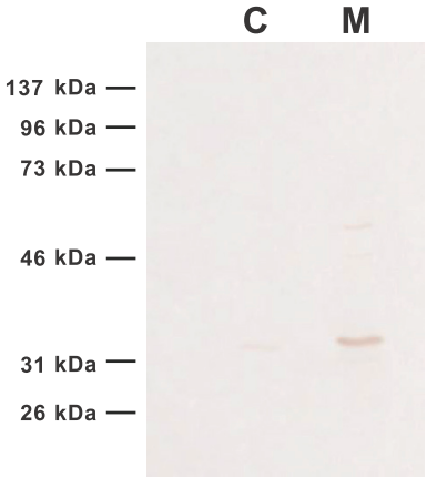
Figure. Western blotting of M3527-isolated cytoplasmic fraction (C) and mitochondria (M)
Mitochondrial proteins were extracted with RIPA Buffer, and cytoplasmic fraction and mitochondria were detected with VDAC1/2 antibody.
M3527 allows for highly efficient purification of mitochondria.
Nuclear / Cytoplasmic Fractionation Kit
Nuclear / Cytoplasmic Fractionation Kit (Product No. N1208) is a product for convenient fractionation of nuclear and cytoplasmic proteins from cultured mammalian cells. By using the three reagents included, the quick collection of each fraction can be achieved. The extracted proteins can be used directly in downstream applications such as Western Blotting.
Kit Components
- 1X Cytoplasmic Extraction Buffer for approx. 50 samples
- 20X Detergent Solution for approx. 50 samples
- 1X Nuclear Extraction Buffer for approx. 50 samples
Application
[for Cytoplasmic Extraction]- Collect cells and wash cells twice with ice cold PBS. Use a cell scraper for adherent cells.
- Resuspend the cells gently in cold 1X Cytoplasmic Extraction Buffer. Add 400 μL of the buffer to 100 μL of the cell volume (~1 x 10⁷ cells).
- Incubate on ice for 10 minutes.
- Add 20x Detergent solution to the cell suspension and vortex vigorously for 10 seconds. Use 25 μL of 20X Detergent Solution per 500 μL of the solution from step 3.
- Centrifuge at 800 x g for 10 minutes at 4°C. Transfer the supernatant (cytoplasmic fraction) to a new tube for downstream experiments.
- Resuspend the pellet in cold 1X Nuclear Extraction Buffer. Add 100 μL of 1X Nuclear Extraction Buffer per 100 μL pellet.
- Incubate the pellet solution on ice for 20 minutes, vortexing every 5 minutes.
- Centrifuge at 15,000 x g for 10 minutes at 4°C.
- Transfer the supernatant (nuclear fraction) to a new tube for downstream experiments.

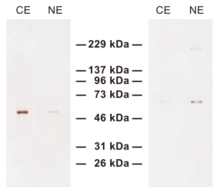
Figure. Western Blotting of a cytoplasmic fraction (CE) and nuclear fraction (NE) extracted with N1208
The fractions were detected using α-Tubulin antibody (left) and Lamin B1 antibody (right). Excellent separation was achieved.

