Make sure to sign up for an account today for exclusive coupons and free ground shipping on orders over $75!
Maximum quantity allowed is 999
Intracellular reactive oxygen species (ROS) are mainly produced in mitochondria. ROS react to lipids or proteins inside cells may inhibit the cell functions and induce a series of pathological processes including cell death, aging and oncogenesis.
The Intracellular Reactive Oxygen Species (ROS) Detection Assay Kit provides a fluorescence probe, DCFH-DA, suitable for immediate detection of ROS. This kit is sufficient for one 96-well microplate assay and requires 1 mL of test solution for 1 × 10⁶ cells/mL. The optimal working concentration of the reaction dye may vary depending on the cell lines being used.
Kit Components
- 1000×Reaction Dye (DCFH-DA) : 10 μL for 100 tests
- 10×Reaction Buffer : 25 mL×2 for 100 tests
Application 1: Fluorescence observation of ROS produced in Hela cells
- Culture the HeLa cells in 96 well culture plate and grow to 80% confluent.
- Remove the culture media. Treat the cells with 1 × Reaction Dye and incubate for 30 minutes in CO2 incubator (37°C).
- Observe the Intracellular ROS (green fluorescence) by fluorescence microscope.
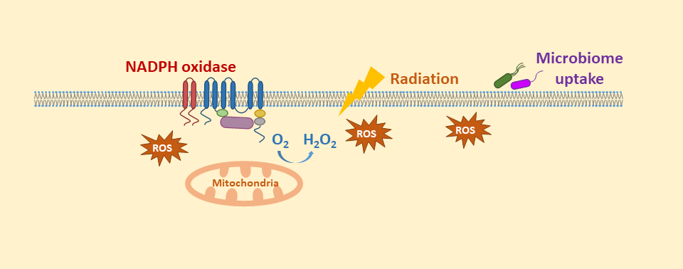
Figure 1. Fluorescence microscopy observation of ROS produced in Hela cells
ROS produced in HeLa cells were observed by fluorescence microscope.
Application 2-1: Comparison of ROS produced in NIH-3T3 treated with a ROS inducer and a ROS inhibitor
- Culture the NIH-3T3 cells in 96 well culture plate and grow to 80% confluent.
- Remove the culture media from a sample of cells and treat cells with a ROS inducer and incubate in a CO2 incubator (37°C) overnight.
- Remove the ROS inducer from a sample of cells and additionally treat cells with a ROS inhibitor and incubate in a CO2 incubator (37°C) for 30 minutes.
- Treat all cells with 1 × Reaction Dye in a CO2 incubator (37°C) for 30 minutes.
- Test intracellular ROS production on a plate reader.
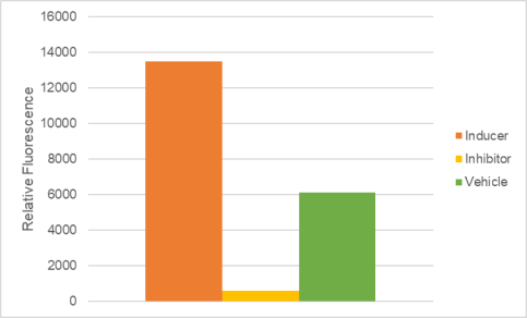
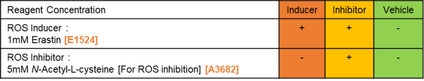
Figure 2. Detection of ROS in NIH-3T3 cells using a plate reader
ROS production was induced by a ROS inducer and inhibited by a ROS inhibitor.
Application 2-2: ROS analyzed in NIH-3T3 with and without a ROS inducer by flow cytometry
- Culture the NIH-3T3 at 105 cells/mL and grow to 80% confluent.
- Remove the culture media from a sample of cells and treat cells with 1 mM Erastin and incubate in a CO2 incubator (37°C) overnight.
- Collect the cells and treat cells with 1 × Reaction Dye in a CO2 incubator (37°C) for 30 minutes.
- Analyze the intracellular ROS production by flow cytometry.
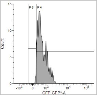
Figure 3. Detection of ROS in NIH-3T3 cells by flow cytometer
ROS production was induced by Erastin treatment.