Caution Notice:
It has come to our notice that certain fraudulent individuals or entities are misusing our Company’s name and TCI’s registered trademarks by promoting and offering for sale regulated and hazardous chemical substances, including Potassium Cyanide, through online platforms like YouTube. We hereby categorically clarify that TCI has no association or connection whatsoever with the products being displayed or sold in these or similar videos. These products have been falsely represented as being associated with TCI, and the unauthorized use of our trademark and brand name is both illegal and misleading. TCI Chemicals is a responsible global organization committed to adhering to all applicable international and domestic regulatory frameworks. We do not manufacture, distribute, or engage in the sale of regulated chemical substances in India or in any other jurisdiction unless specifically authorized by the relevant laws and regulatory agencies. All our operations and offerings are governed by stringent safety, quality, and compliance protocols in accordance with applicable laws. Further, TCI Chemicals markets and sells its products exclusively through its official website and authorized distributors. We do not sell, endorse, or supply our products through any third-party platforms, unauthorized agents, or resellers. Any individual or organization purchasing products outside these official channels does so at their own risk, and TCI disclaims all responsibility and liability for any consequences arising therefrom. Members of the public, customers, and partners are strongly advised to exercise caution and conduct due diligence before engaging in any transactions involving products claiming association with TCI Chemicals. The official list of our authorized distributors is publicly available and may be accessed at: https://www.tcichemicals.com/IN/en/distributor/index. We are currently pursuing appropriate legal remedies against those misusing our brand, and we reserve all rights available to us under applicable intellectual property and criminal laws. If you become aware of any such fraudulent activity or require clarification, you may reach out to us at: Sales-IN@TCIchemicals.com. Your vigilance and cooperation are essential in safeguarding the public interest and protecting the integrity of the TCI brand.
Maximum quantity allowed is 999

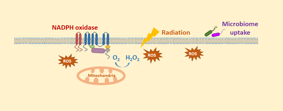
Intracellular reactive oxygen species (ROS) are mainly produced in mitochondria. ROS react to lipids or proteins inside cells may inhibit the cell functions and induce a series of pathological processes including cell death, aging and oncogenesis.
The Intracellular Reactive Oxygen Species (ROS) Detection Assay Kit provides a fluorescence probe, DCFH-DA, suitable for immediate detection of ROS. This kit is sufficient for one 96-well microplate assay and requires 1 mL of test solution for 1 × 10⁶ cells/mL. The optimal working concentration of the reaction dye may vary depending on the cell lines being used.
Kit Components
- 1000×Reaction Dye (DCFH-DA) : 10 μL for 100 tests
- 10×Reaction Buffer : 25 mL×2 for 100 tests
Application 1: Fluorescence observation of ROS produced in Hela cells
- Culture the HeLa cells in 96 well culture plate and grow to 80% confluent.
- Remove the culture media. Treat the cells with 1 × Reaction Dye and incubate for 30 minutes in CO2 incubator (37°C).
- Observe the Intracellular ROS (green fluorescence) by fluorescence microscope.

Figure 1. Fluorescence microscopy observation of ROS produced in Hela cells
ROS produced in HeLa cells were observed by fluorescence microscope.
Application 2-1: Comparison of ROS produced in NIH-3T3 treated with a ROS inducer and a ROS inhibitor
- Culture the NIH-3T3 cells in 96 well culture plate and grow to 80% confluent.
- Remove the culture media from a sample of cells and treat cells with a ROS inducer and incubate in a CO2 incubator (37°C) overnight.
- Remove the ROS inducer from a sample of cells and additionally treat cells with a ROS inhibitor and incubate in a CO2 incubator (37°C) for 30 minutes.
- Treat all cells with 1 × Reaction Dye in a CO2 incubator (37°C) for 30 minutes.
- Test intracellular ROS production on a plate reader.
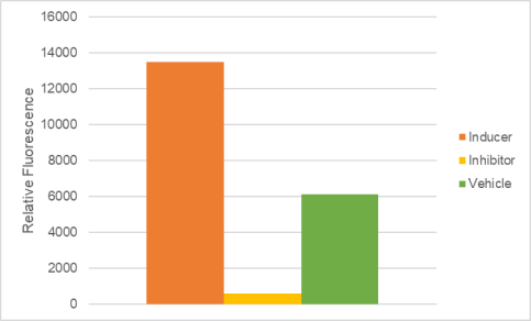

Figure 2. Detection of ROS in NIH-3T3 cells using a plate reader
ROS production was induced by a ROS inducer and inhibited by a ROS inhibitor.
Application 2-2: ROS analyzed in NIH-3T3 with and without a ROS inducer by flow cytometry
- Culture the NIH-3T3 at 105 cells/mL and grow to 80% confluent.
- Remove the culture media from a sample of cells and treat cells with 1 mM Erastin and incubate in a CO2 incubator (37°C) overnight.
- Collect the cells and treat cells with 1 × Reaction Dye in a CO2 incubator (37°C) for 30 minutes.
- Analyze the intracellular ROS production by flow cytometry.
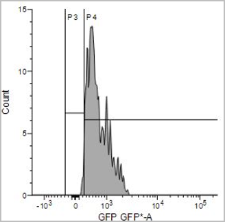
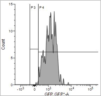
Figure 3. Detection of ROS in NIH-3T3 cells by flow cytometer
ROS production was induced by Erastin treatment.

