Maximum quantity allowed is 999

Antibody-binding proteins such as protein A, protein G and protein L are bacterial proteins that bind specifically to antibodies. They are mainly used for antibody purification, immunoprecipitation (IP) and immunodetection. Each antibody binding protein exhibits different affinities to various animal species and antibody subtype. Therefore, it is important to determine the appropriate antibody-binding protein for your sample of interest. TCI has a wide variety of protein A, protein G and protein L conjugates for you to select from.
Binding Affinity of Protein A, Protein G and Protein L
| Animal Species | Antibody Subtype | Protein A | Protein G | Protein L (*) |
|---|---|---|---|---|
| Human | IgG1 | ++ | ++ | ++ |
| IgG2 | ++ | ++ | ++ | |
| IgG3 | -- | ++ | ++ | |
| IgG4 | ++ | ++ | ++ | |
| IgM | - | -- | ++ | |
| IgD | -- | -- | ++ | |
| IgA | - | -- | ++ | |
| Mouse | IgG1 | ++ | ++ | ++ |
| IgG2a | ++ | ++ | ++ | |
| IgG2b | -- | ++ | ++ | |
| IgG3 | ++ | ++ | ++ | |
| IgM | - | -- | ++ | |
| Rat | IgG1 | - | + | ++ |
| IgG2a | -- | ++ | ++ | |
| IgG2b | -- | + | ++ | |
| IgG2c | ++ | ++ | ++ | |
| Goat | Total IgG | + | ++ | -- |
| Bovine | Total IgG | + | ++ | -- |
| Rabbit | Total IgG | ++ | ++ | - |
| Chicken | Total IgY | -- | -- | -- |
++: Strong; +: Medium; -: Low; --: None
* Protein L binding is restricted to antibodies that contain the right subtypes of κ light chains.
Protein A
Protein A is a bacterial cell wall component from Staphylococcus aureus that specifically binds to the Fc region of IgG derived from various species including human, rabbit, mouse and cow. Our products consist of a recombinant protein A mutant which allows elution of antibodies under mild conditions (pH 4.0). As there is no change in antibody binding affinity, it can be used in the same way as normal protein A.
Products
Application:Purification of human IgG using P2461
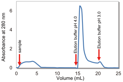
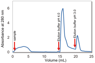
Protein A agarose in which protein A is bound to an agarose resin by a covalent coupling method can be used in antibody purification and immunoprecipitation. Antibody purification using protein A agarose usually requires an acidic buffer solution between pH 2.5 and pH 3.0 during elution steps. However, this frequently causes the antibody to undergo acid denaturation, changing its higher-order structure, resulting in antibody aggregation and inactivation. TCI’s protein A agarose uses a genetically modified protein A mutant which allows for the elution of antibodies under mild conditions (pH 4.0), under which most antibodies do not denature, as shown in Figure 1.
Protocol:
- Fill the column with protein A agarose (Product No. P2461), and equilibrate it with binding buffer.
- Add human IgG.
- Wash the resin with binding buffer, and then elute antibodies with pH 4.0 and pH 3.0 elution buffer.


The majority of applied human IgG was successfully eluted at pH4.0 when using P2461.
Protein G
Protein G is a bacterial cell wall component of Group G Streptococci strain. It binds specifically to the Fc region of immunoglobulins (especially IgG) and weakly to the Fab fragment.
Products
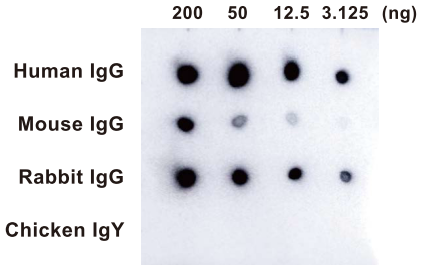
Application:Detection of various IgGs by P2962 using the dot-blot method
Protocol:
- Spot Human IgG, mouse IgG, rabbit IgG, and chicken IgY at a 4-fold dilution from 200 ng onto PVDF membrane.
- Blocked for 60 minutes at room temperature.
- Added Protein G HRP Conjugate (Product No. P2962) and incubate for 60 minutes at room temperature.
- Detect the spots by chemiluminescence.

Protein L
Protein L is a cell wall molecule from the bacterial species Peptostreptococcus magnus. It binds immunoglobulin light chains in a wide range of species including human, mouse, rat, pig, and hamster, and can bind to any immunoglobulin isoform containing a kappa light chain (IgG, IgM, IgA, IgE, and IgD). It can also bind single-chain antibodies (scFv) and Fab fragments with κ light chains.
Products
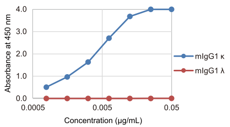
Application:Detection of a κ light chain using P2999
Protocol:
- Coat mouse IgG κ and IgG λ onto ELISA plates.
- Block for 2 hours at room temperature .
- Add Protein L HRP Conjugate (Product No. P2999) and incubate for 30 minutes at room temperature.
- Add TMB solution and incubate for 30 minutes.
- Add 1N HCl solution and measure the absorbance at 450 nm.


