Published TCIMAIL newest issue No.197
Maximum quantity allowed is 999
Please select the quantity
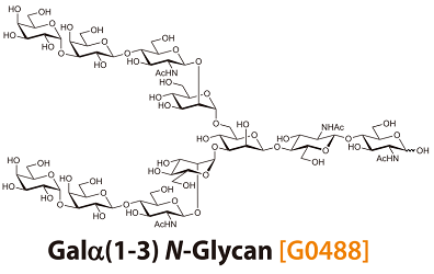
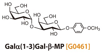
Anti-αGal Polyclonal Antibodies —Antibodies capable of detecting αGal epitope(Galα1-3Gal)—
Anti-αGal antibody exists as a natural antibody in humans. Binding of this antibody to Galαantigens (αGal epitope) expressed on porcine xenograft surfaces are a major factor for determining engraft survival. Recently, it has been observed that therapeutic antibodies and cell processing material for reproductive medicine contain the αGal epitope, which indicates the importance of rapid detection of αGal epitope.
Products
*These antibodies are unavailable in the U.S. and China.
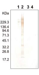
Anti-αGal antibody can be utilized for detection of the αGal epitope on glycoproteins
|
|
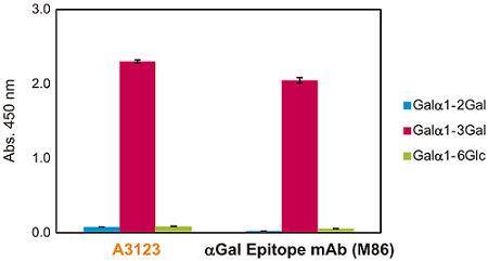
Anti-αGal antibody shows the same high specificity compared with an anti-αGal monoclonal antibody
Secondary Antibodies and Streptavidins
Anti-Chicken IgY
*These antibodies are unavailable in the U.S. and China.


